
Figure 6. Modeled DNA interaction positioned in the NuiA-NucA complex.
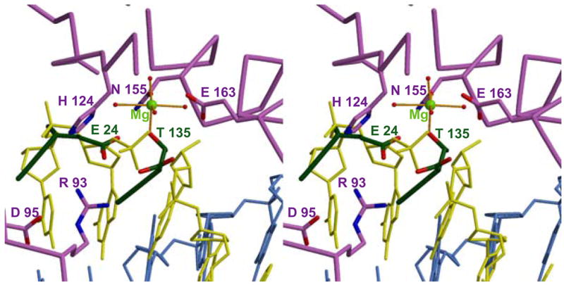
The extent to which NuiA mimics the substrate is illustrated by this stereoview of a docked model of a cleaved DNA octamer in the active site of NucA. Selected residues from NucA and NuiA are indicated in lavender and green, respectively. The model is based on the alignment of active site residues in the NucA structure (Arg122-Ile125 and Met147-Arg156) with active site residues in the structure of Vvn in complex with DNA (Trp78-Val81 and Leu119-Gly128; PDB ID 1OUP). The coordination of the active site Mg2+ (green) to four water molecules as well as to Asn155 (lavender) and to a phosphate oxygen of the cleaved DNA strand (yellow) are indicated. The active site residues are shown in grey and the complementary strand of the DNA is shown in blue.