
Fig. 2.
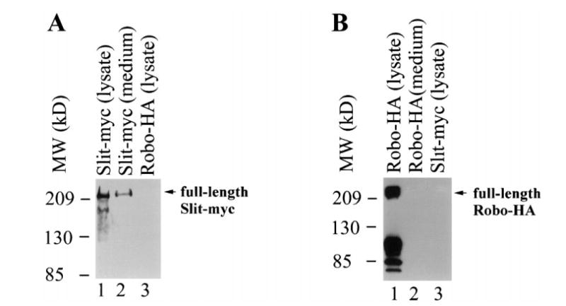
Extracellular secretion of Slit, but not Robo. Shown here are results from western blots. (A) The monoclonal anti-myc antibody detected Slit-myc in both the conditioned medium (lane 1) and lysates (lane 2) of cells transfected with slit-myc, but not the lyate (lane 3) of cells transfected with robo-HA. The upper band is equivalent to the band produced by in vitro translation of an mRNA encoding the full-length Slit-myc. Multiple bands of Slit in addition to the full-length Slit-myc were observed. (B) The monoclonal anti-HA antibody detected Robo-HA in the lysates (lane 1), but not in the medium (lane 2), of cells transfected with robo-HA. It did not recognize Slit-myc (lane 3). The uppermost band is equivalent to the band produced by in vitro translation of an mRNA encoding the full-length Robo-HA. Multiple bands of Robo-HA in addition to the full-length were observed.