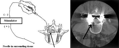
Fig. 2.
To the left: The stimulation technique used to assess pedicle screw placements. The same technique can be used to test markers, taps, and pedicle holes. In this case, the pedicle screw has broken through the wall of the pedicle and is situated very close to an emerging nerve root (marked by arrow). As a result, electrical current, following the path of least resistance through the pedicle screw and the breach in the pedicle wall, is expected to excite the nerve root. Excitation typically occurs at very low stimulation intensities and results in triggered myogenic responses from muscles innervated by the nerve root. To the right: The CT scan showed the pedicle screw has entered the spinal canal and is in a position where L5 spinal nerve root (marked by arrow), was found on surgical inspection to be significantly compromised. (Combined and modified from: Toleikis, JR et al. The usefulness of electrical stimulation for assessing pedicle screw placements. J. Spin. Disord. 2000, 13(4), 283–289 and Toleikis, JR et al. The use of dermatomal evoked responses during surgical procedures that use intrapedicular fixation of the lumbosacral spine. Spine 1993, 18(16), 2401–2407)

