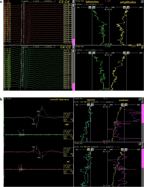
Fig. 2.
a Trend analysis of cerebro-spinal evoked potentials. Left of the picture shows the pathological polyphasic D-waves from the beginning, indicating chronic demyelinisation of the corticospinal tract on both sides. During the operation, as shown on the right, significant alterations of the latencies and amplitudes of the D-waves were observed leading to adaption of the surgical approach. b Trend analysis of neuro-spinal and neuro-cerebral evoked potentials of right and left tibial nerves showing continuous reduction of amplitudes on both sides due to systemic drug, vascular and temperature effects

