
Figure 7.
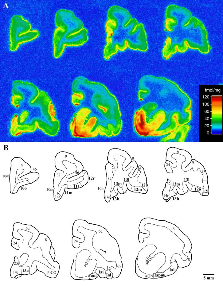
A) Coronal autoradiograms of [3H]cyanoimipramine binding in the prefrontal cortex of the vervet monkey. Sections are arranged in a rostral to caudal manner with the most rostral section located in the upper left corner and the most caudal section located in the lower right corner. Sections are not spaced at regular intervals. The scale bar denotes density of binding (fmol [3H]cyanoimipramine/mg protein). B) Sketches of the autoradiograms shown in A. The 15 orbitofrontal areas for which [3H]cyanoimipramine binding was quantified are shown in bold with architectonic boundaries delineated. Scale bar = 5mm.