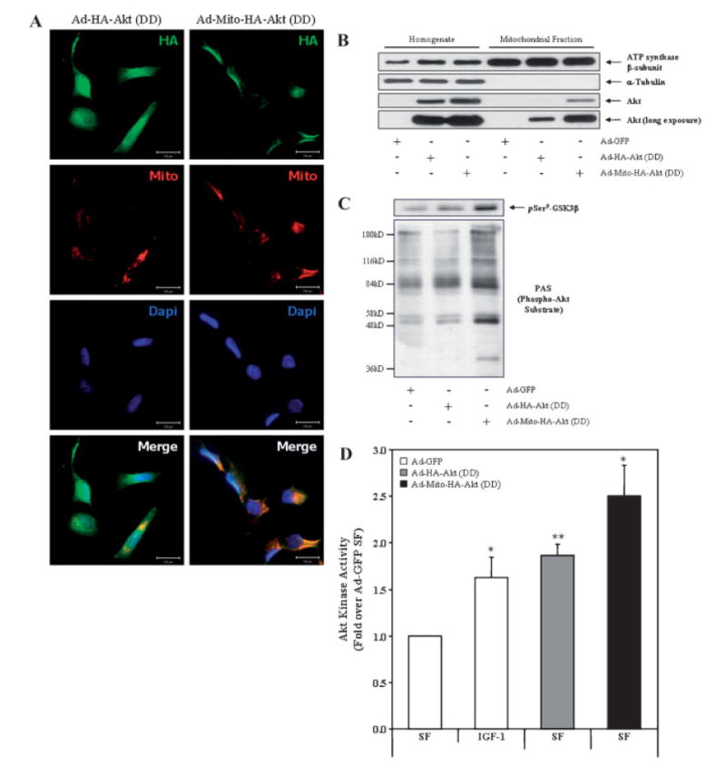
Fig. 4.

Ad-Mito-HA-Akt (DD) colocalizes with mitochondria and exhibits constitutive activation. A: SH-SY5Y cells seeded on chamber sides were infected with Ad-HA-Akt (DD) or Ad-Mito-HA-Akt (DD) and serum starved overnight. Cells were first loaded with Mitotracker Red CM-H2TMRos (Mito; red) to label mitochondria then fixed and labeled with the monoclonal anti-HA antibody (HA; green) to visualize the Akt adenoviral constructs and Dapi (Dapi; blue) to counterstain the nuclei. B: Homogenates and mitochondrial fractions from SH-SY5Y cells infected overnight with the indicated adenoviral constructs were immunoblotted for ATP synthase β-subunit (mitochondrial protein), α-tubulin (cytosolic protein), and Akt. C: SH-SY5Y cells infected with the designated adenoviral constructs were serum starved overnight, mitochondrial fractions prepared and probed for GSK3β phospho-Ser9 (pSer9-GSK3β) and phospho-Akt Substrate (PAS). D: SH-SY5Y cells infected with designated adenoviral constructs were serum starved overnight prior to incubation in the absence (serum free [SF]) or presence (IGF-1) of 10 nM IGF-1 for 15 min followed by preparation of mitochondria. Akt was immunoprecipitated from the mitochondrial fractions, and Akt kinase activity was determined by an in vitro kinase assay. The data are presented as mean ± SEM, n = 5 independent experiments; *, P < 0.05 versus Ad-GFP SF; **, P < 0.005 versus Ad-GFP SF (two-tailed, paired Student’s t-test).
