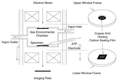
Figure 1.
Schematic diagram of the EC. The upper and lower windows are covered with carbon sealing films held on copper grids with nine apertures. The interior of the EC is constantly circulated with water vapor to keep the specimen placed on the lower carbon film in wet state. The EC contains an ATP-containing microelectrode to apply ATP to the specimen iontophoretically. The image of the specimen is recorded on the imaging plate.

