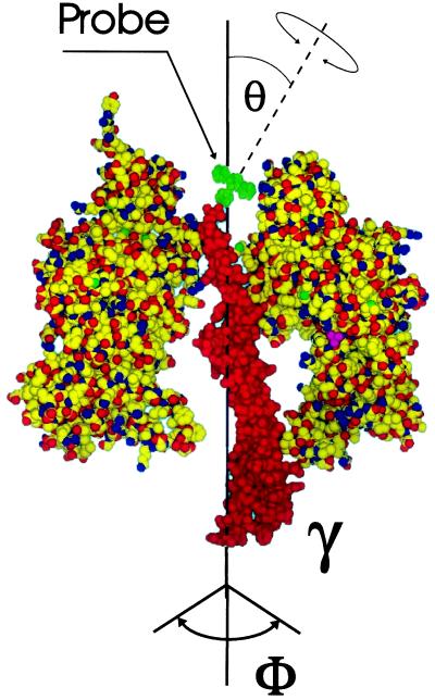
Figure 1.
Model of the partial structure of chloroplast CF1 from spinach shaped after the structure of mitochondrial MF1 (13): the γ-subunit and two subunits of the (αβ)3-hexagon are shown. The dye eosin (green) was bound by its maleimide function to the penultimate residue, Cys-322, of γ (25, 26). Rotational mobilities of γ and eosin are indicated by arrows. θ denotes the effective inclination angle of the dye’s bond axis relative to the long axis of γ.

