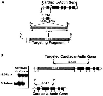
Figure 1.
Targeted disruption of the cardiac α-actin gene. (A) Generation of the targeting fragment. Exon–intron organization of the cardiac α-actin gene is shown at the top. Open boxes represent noncoding exons, while dark boxes represent coding exons. Exons (1–7) are numbered. The XbaI–SphI fragment used in the targeting construct includes 1.5 kb of sequence upstream and 0.7 kb of sequence downstream of the unique NaeI site. The NaeI site was converted to a SalI site, and the targeting construct was obtained by the ligation of a HPRT minigene SalI cassette. Since the HPRT minigene carries an XbaI site (not shown) the targeting fragment used for electroporation was released from the targeting construct by digestion with KpnI and SphI. The KpnI site is located in the vector (pUC 19). (B) Southern blot pattern of +/+, +/−, and −/− mice. Homologous recombination will lead to the generation of a 5.5-kb band, while the normal allele will give rise to a 3.5-kb band. The illustration to the right shows the location of the PvuII sites in the native and targeted gene. Bar indicates location of the probe used for Southern hybridization. K, KpnI; N, NaeI; S, SphI; Sa, SalI; V, PvuII; and X, XbaI.

