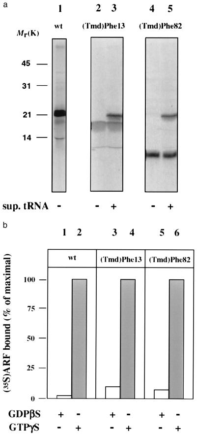
Figure 2.
Characterization of incorporation of (Tmd)Phe into ARF protein. (a) Autoradiograph of an SDS/polyacrylamide gel of [35S]methionine-labeled products from in vitro translations of wild-type (wt) ARF1 (lane 1), ARF-(Tmd)Phe-13 (lanes 2 and 3), and ARF-(Tmd)Phe-82 (lanes 4 and 5) both in the absence (lanes 2 and 4) and presence (lanes 3 and 5) of suppressor tRNA; (b) in vitro-translated wt-ARF (lane 1 and 2), ARF-(Tmd)Phe-13 (lane 3 and 4), and ARF-(Tmd)Phe-82 (lane 5 and 6) were incubated with isolated Golgi membranes from Chinese hamster ovary (CHO) cells in the presence of GDP[βS] (lanes 1, 3, and 5) or GTP[γS] (lanes 2, 4 and 6). After incubation, the amount of radioactivity associated with the membranes was determined.

