Figure 3.
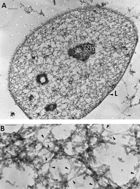
The ultrastructure of the nuclear matrix revealed by resinless section electron microscopy. The nuclear matrix of a CaSki cell was prepared by the cross-link stabilized nuclear matrix preparation procedure and visualized by resinless section electron microscopy (27–29). (A) The nuclear matrix consisted of two parts, the nuclear lamina (L) and a network of intricately structured fibers connected to the lamina and well distributed through the nuclear volume. The matrices of nucleoli (Nu) remained and were connected to the fibers of the internal nuclear matrix. Three remnant nucleoli may be seen in this section. Few intermediate filaments were connected to the outside of the lamina. (B) Seen at higher magnification the highly structured fibers of the internal nuclear matrix seemed to be built on an underlying structure of 10-nm filaments that are occasionally branched. These were seen most clearly when, for short stretches, they were free of covering material (arrowheads). The irregular fibers, with granules well integrated into their structure, may be built on this filamentous core structure. [Bars = 1 μM (A) and 100 nm (B).]

