Abstract
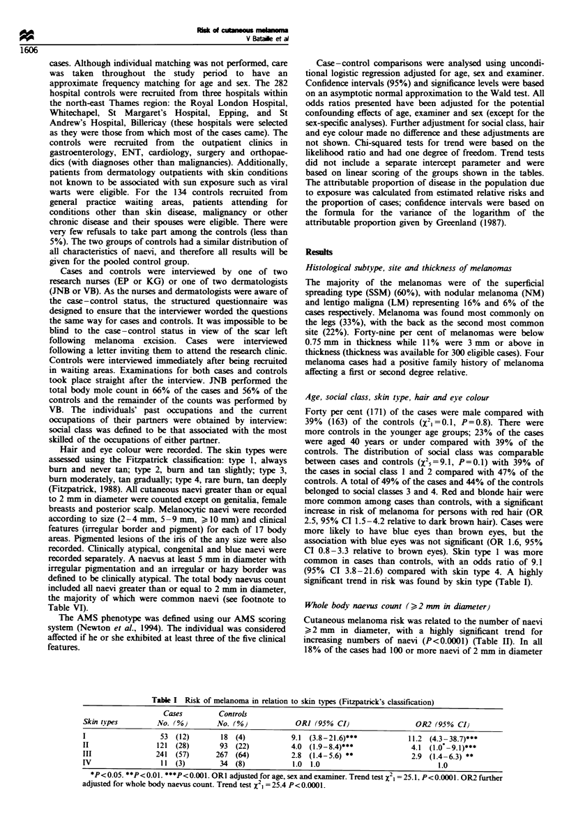
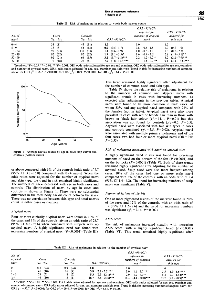
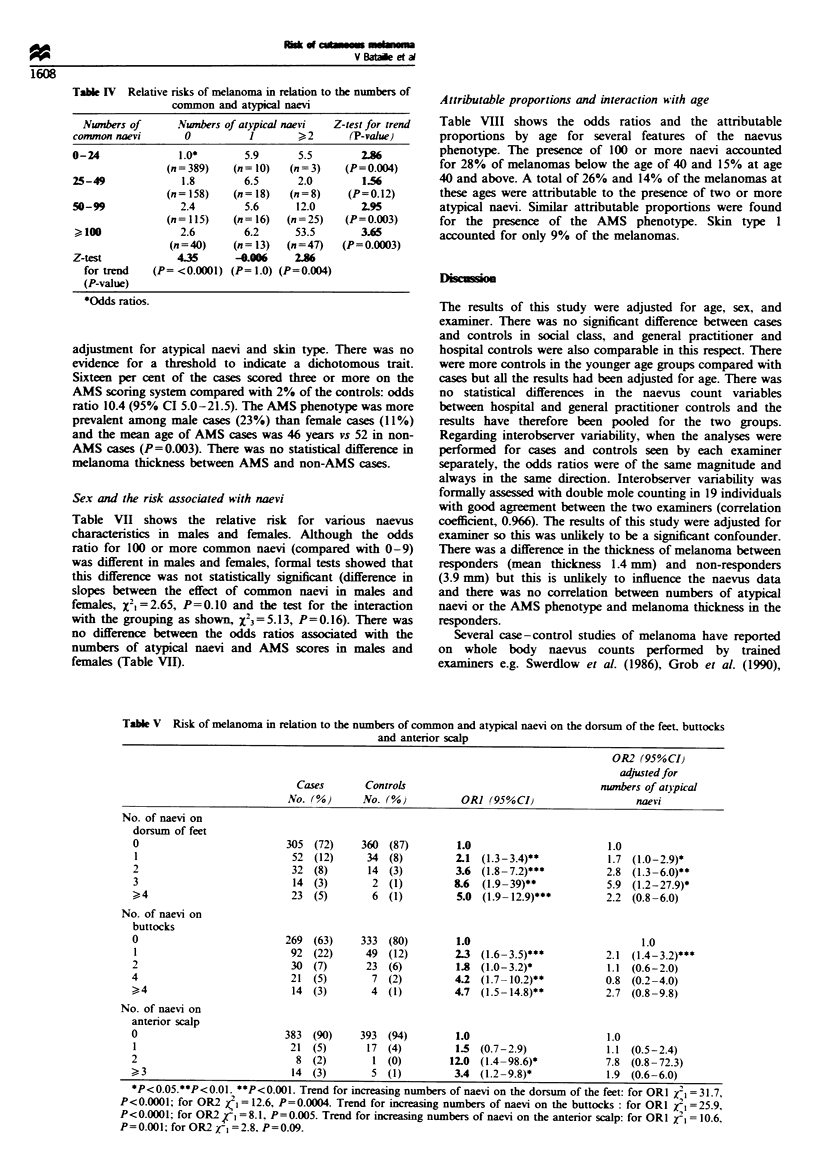
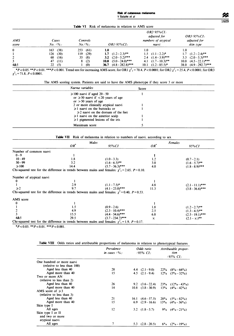
The atypical mole syndrome (AMS) phenotype, characterised by a large number of common naevi as well as atypical naevi, has been described in families with a genetic susceptibility to melanoma. However, the importance of this phenotype for melanoma in the general population has not been conclusively determined. This study was designed to examine the types and distribution of naevi as well as the prevalence of the AMS phenotype in melanoma patients in England compared with controls. A total of 426 cutaneous melanoma cases (61% of all incident cases) aged 16-75 years were recruited between 1989 and 1993 from the north-east Thames region of the UK and 416 controls from the same age group were recruited over the same period and from the same region. Each subject answered a questionnaire covering demographic details, sun exposure history and other risk factors and underwent a skin examination with total body naevus count performed by a dermatologist. The AMS phenotype was defined using a scoring system. Atypical naevi gave the highest relative risk for cutaneous melanoma, with an odds ratio (OR) of 28.7 (P < 0.0001) for four or more atypical naevi compared with none. Many common naevi were also an important risk factor: the OR for 100 or more naevi 2 mm or above in diameter compared with 0-4 naevi was 7.7 (P < 0.0001). Melanoma was also associated with naevi on sun-exposed sites but also with naevi on non-sun-exposed sites such as the dorsum of the feet, buttocks and anterior scalp. Sixteen per cent of the cases had the AMS phenotype compared with 2% of the controls (OR 10.4, P < 0.0001). The AMS phenotype was more common in males than females (P = 0.008). The odds ratio for the presence of the AMS phenotype was dependent on age, with an odds ratio of 16.1 (95% CI 4.6-57.5) for the presence of the AMS phenotype if aged less than 40 compared with an odds ratio of 6.9 (95% CI 2.9-16.6) if aged 40 or more. The AMS phenotype was strongly predictive of an increased risk of melanoma outside the familial context.
Full text
PDF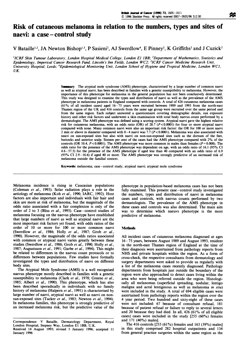
Selected References
These references are in PubMed. This may not be the complete list of references from this article.
- Albert D. M., Searl S. S., Forget B., Lavin P. T., Kirkwood J., Nordlund J. J. Uveal findings in patients with cutaneous melanoma. Am J Ophthalmol. 1983 Apr;95(4):474–479. doi: 10.1016/0002-9394(83)90267-2. [DOI] [PubMed] [Google Scholar]
- Albert L. S., Rhodes A. R., Sober A. J. Dysplastic melanocytic nevi and cutaneous melanoma: markers of increased melanoma risk for affected persons and blood relatives. J Am Acad Dermatol. 1990 Jan;22(1):69–75. doi: 10.1016/0190-9622(90)70010-f. [DOI] [PubMed] [Google Scholar]
- Augustsson A., Stierner U., Rosdahl I., Suurküla M. Common and dysplastic naevi as risk factors for cutaneous malignant melanoma in a Swedish population. Acta Derm Venereol. 1991;71(6):518–524. [PubMed] [Google Scholar]
- Bliss J. M., Ford D., Swerdlow A. J., Armstrong B. K., Cristofolini M., Elwood J. M., Green A., Holly E. A., Mack T., MacKie R. M. Risk of cutaneous melanoma associated with pigmentation characteristics and freckling: systematic overview of 10 case-control studies. The International Melanoma Analysis Group (IMAGE). Int J Cancer. 1995 Aug 9;62(4):367–376. doi: 10.1002/ijc.2910620402. [DOI] [PubMed] [Google Scholar]
- Clark W. H., Jr, Reimer R. R., Greene M., Ainsworth A. M., Mastrangelo M. J. Origin of familial malignant melanomas from heritable melanocytic lesions. 'The B-K mole syndrome'. Arch Dermatol. 1978 May;114(5):732–738. [PubMed] [Google Scholar]
- Fitzpatrick T. B. The validity and practicality of sun-reactive skin types I through VI. Arch Dermatol. 1988 Jun;124(6):869–871. doi: 10.1001/archderm.124.6.869. [DOI] [PubMed] [Google Scholar]
- Gallagher R. P., Elwood J. M., Hill G. B. Risk factors for cutaneous malignant melanoma: the Western Canada Melanoma Study. Recent Results Cancer Res. 1986;102:38–55. doi: 10.1007/978-3-642-82641-2_4. [DOI] [PubMed] [Google Scholar]
- Garbe C., Büttner P., Weiss J., Soyer H. P., Stocker U., Krüger S., Roser M., Weckbecker J., Panizzon R., Bahmer F. Risk factors for developing cutaneous melanoma and criteria for identifying persons at risk: multicenter case-control study of the Central Malignant Melanoma Registry of the German Dermatological Society. J Invest Dermatol. 1994 May;102(5):695–699. doi: 10.1111/1523-1747.ep12374280. [DOI] [PubMed] [Google Scholar]
- Greene M. H., Clark W. H., Jr, Tucker M. A., Kraemer K. H., Elder D. E., Fraser M. C. High risk of malignant melanoma in melanoma-prone families with dysplastic nevi. Ann Intern Med. 1985 Apr;102(4):458–465. doi: 10.7326/0003-4819-102-4-458. [DOI] [PubMed] [Google Scholar]
- Greenland S. Variance estimators for attributable fraction estimates consistent in both large strata and sparse data. Stat Med. 1987 Sep;6(6):701–708. doi: 10.1002/sim.4780060607. [DOI] [PubMed] [Google Scholar]
- Grob J. J., Gouvernet J., Aymar D., Mostaque A., Romano M. H., Collet A. M., Noe M. C., Diconstanzo M. P., Bonerandi J. J. Count of benign melanocytic nevi as a major indicator of risk for nonfamilial nodular and superficial spreading melanoma. Cancer. 1990 Jul 15;66(2):387–395. doi: 10.1002/1097-0142(19900715)66:2<387::aid-cncr2820660232>3.0.co;2-j. [DOI] [PubMed] [Google Scholar]
- Halpern A. C., Guerry D., 4th, Elder D. E., Clark W. H., Jr, Synnestvedt M., Norman S., Ayerle R. Dysplastic nevi as risk markers of sporadic (nonfamilial) melanoma. A case-control study. Arch Dermatol. 1991 Jul;127(7):995–999. [PubMed] [Google Scholar]
- Halpern A. C., Guerry D., 4th, Elder D. E., Trock B., Synnestvedt M. A cohort study of melanoma in patients with dysplastic nevi. J Invest Dermatol. 1993 Mar;100(3):346S–349S. doi: 10.1111/1523-1747.ep12470256. [DOI] [PubMed] [Google Scholar]
- Holly E. A., Kelly J. W., Shpall S. N., Chiu S. H. Number of melanocytic nevi as a major risk factor for malignant melanoma. J Am Acad Dermatol. 1987 Sep;17(3):459–468. doi: 10.1016/s0190-9622(87)70230-8. [DOI] [PubMed] [Google Scholar]
- Koh H. K., Kligler B. E., Lew R. A. Sunlight and cutaneous malignant melanoma: evidence for and against causation. Photochem Photobiol. 1990 Jun;51(6):765–779. [PubMed] [Google Scholar]
- Newton Bishop J. A., Bataille V., Pinney E., Bishop D. T. Family studies in melanoma: identification of the atypical mole syndrome (AMS) phenotype. Melanoma Res. 1994 Aug;4(4):199–206. doi: 10.1097/00008390-199408000-00001. [DOI] [PubMed] [Google Scholar]
- Newton J. A., Bataille V., Griffiths K., Squire J. M., Sasieni P., Cuzick J., Bishop D. T., Swerdlow A. How common is the atypical mole syndrome phenotype in apparently sporadic melanoma? J Am Acad Dermatol. 1993 Dec;29(6):989–996. doi: 10.1016/0190-9622(93)70279-3. [DOI] [PubMed] [Google Scholar]
- Nordlund J. J., Kirkwood J., Forget B. M., Scheibner A., Albert D. M., Lerner E., Milton G. W. Demographic study of clinically atypical (dysplastic) nevi in patients with melanoma and comparison subjects. Cancer Res. 1985 Apr;45(4):1855–1861. [PubMed] [Google Scholar]
- Rodriguez-Sains R. S. Ocular findings in patients with dysplastic nevus syndrome. Ophthalmology. 1986 May;93(5):661–665. doi: 10.1016/s0161-6420(86)33684-4. [DOI] [PubMed] [Google Scholar]
- Swerdlow A. J., English J., MacKie R. M., O'Doherty C. J., Hunter J. A., Clark J., Hole D. J. Benign melanocytic naevi as a risk factor for malignant melanoma. Br Med J (Clin Res Ed) 1986 Jun 14;292(6535):1555–1559. doi: 10.1136/bmj.292.6535.1555. [DOI] [PMC free article] [PubMed] [Google Scholar]
- Traupe H., Macher E., Hamm H., Happle R. Mutation rate estimates are not compatible with autosomal dominant inheritance of the dysplastic nevus "syndrome". Am J Med Genet. 1989 Feb;32(2):155–157. doi: 10.1002/ajmg.1320320203. [DOI] [PubMed] [Google Scholar]
- Tucker M. A., Greene M. H., Clark W. H., Jr, Kraemer K. H., Fraser M. C., Elder D. E. Dysplastic nevi on the scalp of prepubertal children from melanoma-prone families. J Pediatr. 1983 Jul;103(1):65–69. doi: 10.1016/s0022-3476(83)80777-x. [DOI] [PubMed] [Google Scholar]
- Weinstock M. A., Stryker W. S., Stampfer M. J., Lew R. A., Willett W. C., Sober A. J. Sunlight and dysplastic nevus risk. Results of a clinic-based case-control study. Cancer. 1991 Mar 15;67(6):1701–1706. doi: 10.1002/1097-0142(19910315)67:6<1701::aid-cncr2820670637>3.0.co;2-x. [DOI] [PubMed] [Google Scholar]


