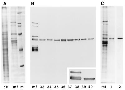
Figure 1.
Purification of MAP60. (A) Protein profiles of carrot cytoskeleton extract (ce) and whole MAP fraction (mf); marker proteins (m) are 205, 116, 97, 68, and 45 kDa. (B) MAPs were fractionated on an FPLC anion exchange column with a linear salt gradient. Individual fractions were analyzed by immunoblotting with whole anti-MAP serum. Fractions 33–40 (146–210 mM salt) were found to contain a protein belonging to the 65-kDa MAPs. The Inset shows this protein to be the 60-kDa MAP. (C) Fractions 36 and 37 (170–186 mM salt) were pooled and further analyzed. Only one band is visible after silver staining on gel (lane 1). Immunoblotting with antibodies against the 65-kDa MAPs confirms the presence of only one protein (lane 2).

