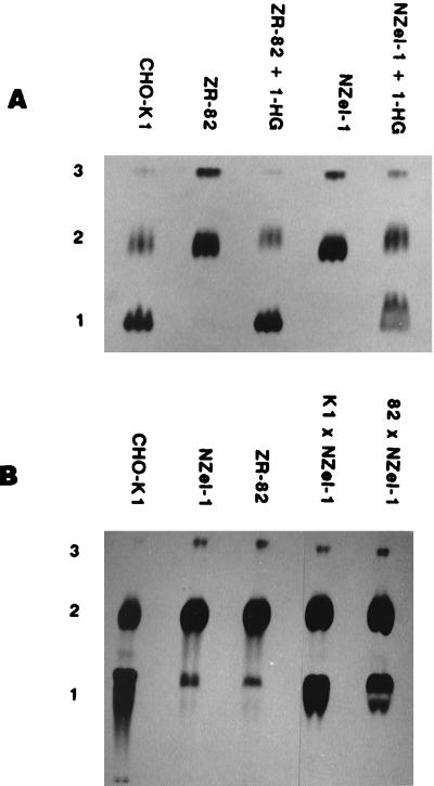
Figure 2.
Subspecies patterns of ethanolamine phospholipids in wild-type cells, mutants, and fusates. Cells (2.5 × 10⁵) were grown for 18 h in sterile glass scintillation vials at 37°C, in medium containing [1-3H]ethanolamine (2 μCi/ml) with or without 20 μM 1-hexadecylglycerol (1-HG) (A). The medium was removed, the cells were washed once with 2 ml PBS, and the lipids were extracted in 3.8 ml CHCl3/methanol/PBS (1:2:0.8) containing 200 μg of a carrier lipid (beef brain PE). After transfer to test tubes, 1 ml CHCl3 and 1 ml PBS were added to form the two-phase Bligh and Dyer system (19) and the lower (organic) phase was collected after centrifugation. Solvent was removed using a stream of nitrogen and the labeled phospholipids were separated by two-stage single-dimension TLC (14). The labeled species were visualized by autoradiography at −80°C after spraying the plates with EN3HANCE. Hybrid cell lines (B) were generated as described. The first cell line listed in the hybrid pairings were the lines that carried the secondary mutations required for hybrid selection. Band 1, plasmenylethanolamine (Rf = 0.3); band 2, PE (Rf = 0.5); band 3, unknown (Rf = 0.9).

