
Figure 1. Developmental defects during the formation of Meckel’s cartilage and the mandible bone in Tgfbr2fl/fl;Wnt1-Cre mice.
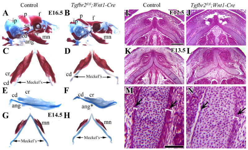
(A,B) Lateral view of skeletal staining preparations of control and Tgfbr2fl/fl;Wnt1-Cre mutant mice at E16.5. Tgfbr2fl/fl;Wnt1-Cre mice have severe defects of the frontal (f), parietal (p) and mandible bones (mn). (C,D) Top view of Meckel’s cartilage and the mandible bone at E16.5 in control and Tgfbr2fl/fl;Wnt1-Cre mutant mice. (E,F) Lateral view of mandible complexes stained with Alcian Blue at E16.5 in control and Tgfbr2fl/fl;Wnt1-Cre mutant mice. Tgfbr2fl/fl;Wnt1-Cre mice have a shortened mandible bone, diminished coronoid and condylar process, and absent angular process. (G,H) Top view of Meckel’s cartilage and the mandible bone at E14.5 in control and Tgfbr2fl/fl;Wnt1-Cre mutant mice samples. (I–M) Histological analysis of Meckel’s cartilage in transversal sections at E12.5 (I,J) and E13.5 (K–N). At E12.5, the Meckel’s cartilage in the Tgfbr2fl/fl;Wnt1-Cre mutant sample is indistinguishable from control. At E13.5, the Tgfbr2fl/fl;Wnt1-Cre mutant sample has defects including a curved Meckel’s cartilage and disrupted layers of the perichondrium and alignment of the chondrocytes in Meckel’s cartilage (arrows). The white parentheses indicate the thickness of the layers. Scale bars: 200μm in I–L; 100μm in M, N.
f; frontal bone, p; parietal bone, ip; interparietal bone, sq; squamous bone, mn; mandible bone, cd; condylar process, cr; coronoid process, ang; angular process, Meckel’s; Meckel’s cartilage