
Figure 8. CTGF is a downstream mediator of TGF-β signaling to control cell proliferation in Meckel’s cartilage.
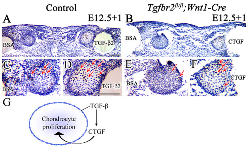
BrdU labeling of cultured Meckel’s cartilage from control and Tgfbr2fl/fl;Wnt1-Cre mice at E12.5, treated with BSA, TGF-β2 or CFGF beads for 24 hours (E12.5+1). (A,C,D) Control sample treated with BSA and TGF-β2 beads. The number of BrdU labeled cells (red arrows) is much greater in Meckel’s cartilage treated with TGF-β2 beads. (B,E,F) Tgfbr2fl/fl;Wnt1-Cre sample treated with BSA and CTGF beads. The Tgfbr2fl/fl;Wnt1-Cre sample has few labeled cells with BSA beads but many more after treatment with CTGF beads (red arrows). (G) Schematic drawing demonstrates that TGF-β controls CTGF expression in the perichondrium to regulate chondrocyte proliferation during Meckel's cartilage development. Scale bar: 100μm in A–F.