
Figure 9. TGF-β signaling is required for proper development of the condylar process.
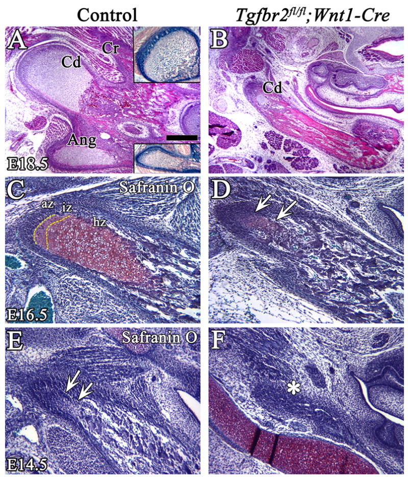
Histological analysis of the coronoid, condylar and angular processes in control and Tgfbr2fl/fl;Wnt1-Cre mice. (A,B) H&E staining shows clear zones of endochondral ossification in the condylar and angular processes in the control at E18.5. The insets show X-gal staining of the condyle and angular processes. The endochondral ossification of the condylar process is diminished in the Tgfbr2fl/fl;Wnt1-Cre mutant at E18.5. (C–F) Safranin O staining of the condyle process at E16.5 (C,D) and at E14.5 (E,F). At E16.5, the three zones, articular, intermediate, and hypertrophic, are visible in the control, but chondrogenesis of the condylar process is dramatically diminished and the hypertrophic zone is not detectable in the mutant (arrows). At E14.5, the condylar cartilage matrix is visible in control (arrows), but not in the Tgfbr2fl/fl;Wnt1-Cre mice (asterisk). Scale bar: 200μm in A–H. Cd; condylar process, Cr; coronoid process, Ang; angular process, az; articular zone, iz; intermediate zone, hz; hypertrophic zone.