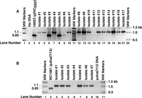
FIG. 1.
Construction of a ΔdnaC::cat mutant. E. coli HME5 carrying the dnaC plasmid, pINCSSD, was transformed as described in Materials and Methods with a DNA fragment encoding the cat gene, which is flanked by DNA that is homologous to the 5′ and 3′ ends of the dnaC gene. PCR analysis of genomic DNA isolated from individual chloramphenicol-resistant colonies was performed with oligonucleotide primers that are complementary to sequences in yjjA and dnaT that border the chromosomal dnaC locus. In panel A, the lanes at the extreme left and right contain DNA fragments whose sizes are indicated at the right of the figure. Lane 11 contains DNA fragments of 1.5, 1.2, 1.0, 0.9, 0.8, 0.7, 0.6, and 0.5 kb. A PCR lacking bacterial DNA corresponds to lane 2. Lanes 3 to 10 and 12 to 21 contain bacterial DNA from HME5 carrying pINCSSD or from the indicated chloramphenicol-resistant recombinants that also contain the dnaC plasmid. The estimated sizes of the amplified DNA are indicated at the left of the figure. In panel B, the ΔdnaC::cat mutation was transferred into E. coli MC1061(pdnaC113) by P1 transduction, using isolate 4 in panel A as the donor strain, followed by selection for chloramphenicol-resistant transductants. PCR analysis was performed as described for panel A either with bacterial DNA isolated from seven independent transductants, with bacterial DNA isolated from MC1061 carrying pdnaC113 (lane 2), or with pdnaC113 DNA (25 ng; lane 10). Lanes 1 and 10 contain DNA fragments whose sizes are indicated at the right of the figure. The estimated sizes of the amplified DNA are noted at the left.

