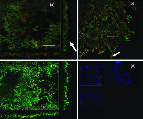
FIG. 3.
Confocal images of biofilms. Scale bars = 75 μm. (a) S. oralis J22 grown in a flow chamber stained with LIVE/DEAD BacLight bacterial viability stain. The arrow indicates carpet-like structures. (b) E. faecalis BS1037 grown on PMMA stained with LIVE/DEAD BacLight bacterial viability stain. The arrow indicates mushroom-like structures. (c) C. albicans SC5314 grown on PMMA stained with FUN1 yeast viability stain. (d) C. albicans SC5314 grown on PMMA stained with calcofluor white, demonstrating the heterogeneous spatial distribution of the biofilms in the x-z and y-z directions and the limited stain penetration in yeast biofilms.

