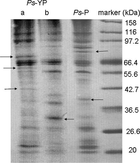
FIG. 1.
Surface protein profiles of YP-grown P. spongiae (Ps-YP) and P-grown P. spongiae (Ps-P) as analyzed by sodium dodecyl sulfate-polyacrylamide gel electrophoresis. Lane a, the surface protein profile after attachment; lane b, surface protein profile before attachment. Samples were analyzed in a 12% separation gel and stained with Coomassie blue. Arrows indicate the unique bands in each sample.

