FIG. 2.
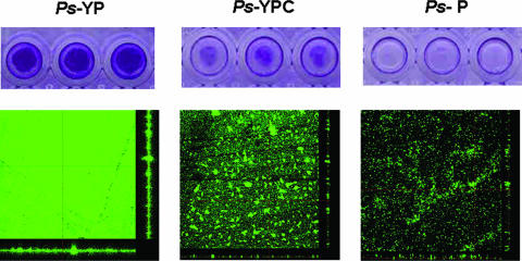
Biofilm formation of Pseudoalteromonas spongiae under different conditions. (Top panels) Biofilm formation analysis in 96-well polystyrene plates with three replicates. The extent of biofilm formation was determined by a crystal violet assay. (Bottom panels) CLSM images of biofilms. Biofilms were stained with fluorescein isothiocyanate-conjugated concanavalin A and were viewed at a ×400 magnification. Ps-YP, Pseudoalteromonas spongiae grown in medium containing yeast extract and peptone; Ps-YPC, YP-grown P. spongiae treated with chloramphenicol at the onset of biofilm formation; Ps-P, Pseudoalteromonas spongiae grown in medium containing peptone.

