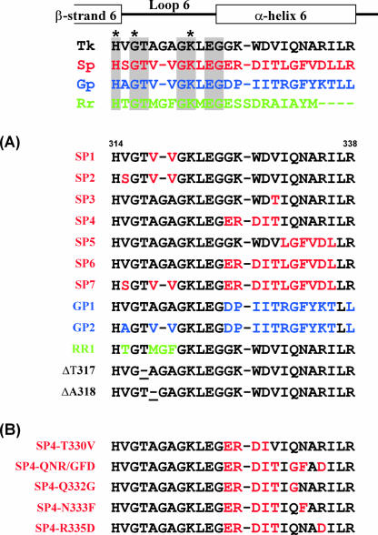
FIG. 1.
Mutant design of RbcTk. Shown is an alignment of the loop 6 and α-helix 6 regions of Rubisco proteins from T. kodakaraensis KOD1 (Tk) (type III; GenBank accession number BAD86479), spinach (Sp) (type I; accession number P00875) (red), G. partita (Gp) (type I; accession number BAA75796) (blue), and R. rubrum (Rr) (type II; accession number P04718) (green). Active-site residues are indicated with asterisks. Residues identical in all four enzymes are shaded. Gaps in the alignment are indicated with hyphens. Secondary structural elements are shown above the alignment. (A and B) Sequences of mutant RbcTk proteins produced in this study. Exchanged residues are indicated with the colors of the sequence on which the mutations were based. The positions where single residues were deleted in ΔT317 and ΔA318 are underlined. (B) Sequences of mutant proteins based on SP4.

