
FIG. 1.
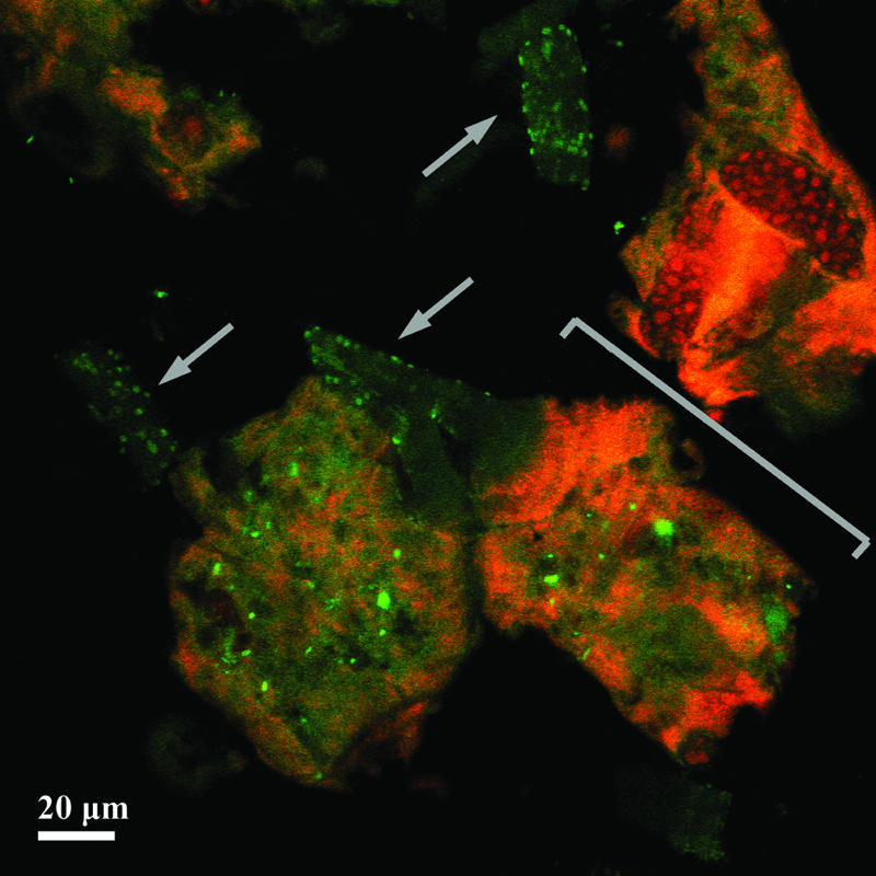
Probe-related signals after FISH applied on a single-granule microsection of 30-μm thickness. The granule was developed in malthouse-derived wastewater. Images are recorded by CLSM after FISH with the Bacteria-specific mix of the EUB338 probes (fluorescein-labeled; green) and the Eukarya-specific probe EUK516 (Cy3-labeled; red). One stack out of 36 stacks of 0.42-μm thickness (each) is shown. Arrows show the branched stalks of a colony of the peritrichous ciliate Epistylis sp. covered by bacteria. The bracket marks the truncated cell bodies (zooids) of the colony.