
FIG. 2.
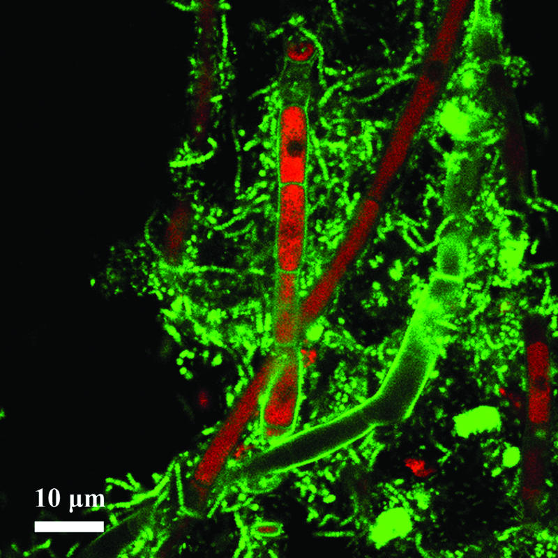
Probe-related signals after FISH applied on a single-granule microsection of 30-μm thickness. The image shows fungal filaments colonized by bacteria in a granule developed in synthetic wastewater. Probe-related signals are recorded by CLSM after FISH with the Bacteria-specific mix of the EUB338 probes (fluorescein-labeled; green) and the Eukarya-specific probe EUK516.