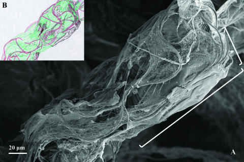
FIG. 5.
Already-dead ciliates from a SBR operated with wastewater released from a brewery. Zooids (cell bodies) and stalks of two adjacent ciliate cells are completely overgrown by bacterial filaments. SEM. Brackets mark the approximate zooid boundaries of the ciliate cell on the right side (A). The scheme (B) redrawn from panel A emphasizes the boundaries between the microorganisms as ciliate cells are colored in black, covered by bacterial filaments (red) and EPS (green).

