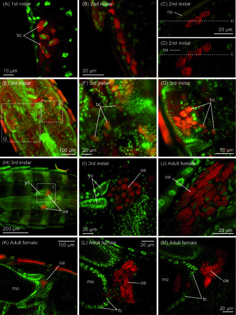
FIG. 4.
Localization of the symbiont in nymphs and adults of C. columbae. (A) Aggregate of bacteriocytes in the lateral body cavity of a first-instar nymph. Cytoplasm of oval bacteriocytes is full of rod-shaped symbiont cells. (B) Aggregate of bacteriocytes in the lateral body cavity of a second-instar nymph. The number of bacteriocytes increases, and the area occupied by the aggregate stretches longitudinally. (C) Distribution of bacteriocytes beneath the hypodermis of the abdominal segment in a first-instar nymph. (D) z-axis reconstruction of the distribution of bacteriocytes through plane D shown in panel C. (E) Migration process of bacteriocytes in a third-instar female nymph. (F and G) Enlarged images of the migrating bacteriocytes. (H) Localization of bacteriocytes in lateral oviducts and formation of ovarial ampullae in a third-instar female nymph. (I) Enlarged image of an ovarial ampulla of the third-instar female nymph. (J) Enlarged image of an ovarial ampulla of a female adult. (K) Ovarial ampulla associated with a mature oocyte in an ovariole. (L) Infection process of symbiont cells from an ovarial ampulla to the posterior pole of an oocyte through follicle cells. (M) Symbiont cells localized in the posterior region of an oocyte. Abbreviations: bc, bacteriocytes; cu, cuticle; fc, follicle cells; mo, mature oocyte; oa, ovarial ampullae; yo, young oocyte.

