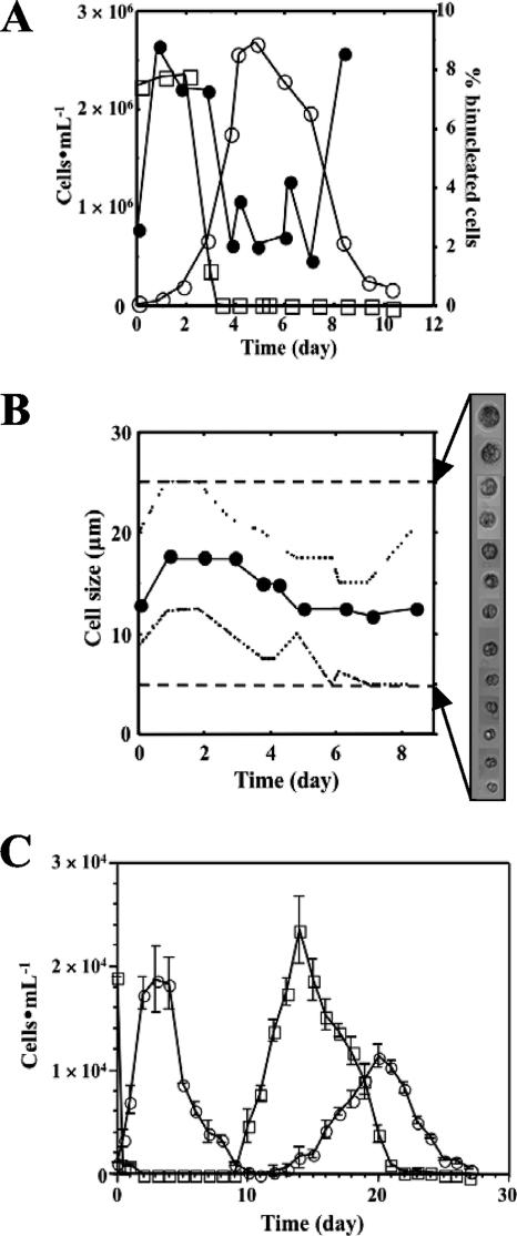
FIG. 1.
Characterization of P. piscicida on algal prey in vitro. (A) Proliferation profile (○) and percentages of binucleated cells (•) for P. piscicida dinospores maintained on Rhodomonas (□) in f/2 medium at 23°C under a cycle of 14 h of light/10 h of darkness. Cells were counted in a hemocytometer after fixation with 4% (wt/vol) formaldehyde in PBS or staining with DAPI (10 μg ml−1) in PBS containing 2.5% (wt/vol) glutaraldehyde as the final concentration. (B) Size distribution of P. piscicida dinospores in standard culture. Shown are the ranges (broken lines) and averages (circles) of P. piscicida dinospore sizes throughout the 8 days of cultivation on Rhodomonas. Cell sizes were measured on digital microscope images (n, 50 cells per sampling). Dashed lines represent the upper and lower limits of detection. (C) Secondary wave of P. piscicida dinospore proliferation in vitro. Cultures were continuously maintained at 23°C with 14 h of light/10 h of darkness over 28 days without additional prey and fresh medium, and P. piscicida (○) and Rhodomonas (□) cell densities were assessed daily (error bars, standard deviations; n, 3 replicate experiments).

