
Figure 2.
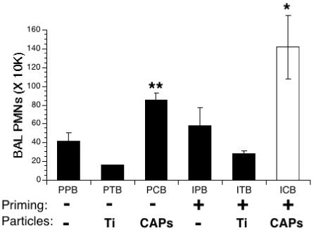
Lung inflammation in IFN-γ-primed, particle-exposed, and S. pneumoniae-infected mice. Mice were primed with IFN-γ (I) or PBS (P) aerosols for 15 min. 3 hours later the same mice were exposed to intranasal solutions of PBS (P), TiO2 (T) or CAPs (C). 24 hours later these mice were infected intranasally with S. pneumoniae (B). Inflammation in the lungs was assessed by counting the number of PMNs in the BAL fluid 24 hours after infection. Data represent the mean ± SEM of at least 3 independent experiments, * = p<0.05 compared to all other groups; **= p<0.03 compared to PPB and PTB groups.