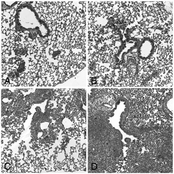
Figure 4.
Histopathology after S. pneumoniae infection with and without antecedent priming and particle exposure. Panels A and B: Little or no acute inflammation (PMN influx) was seen 24 hours after S. pneumoniae infection in control animals (PBS only before infection, PPB, Panel A, and IFN-γ only before infection, IPB, Panel B). Panels C and D: Moderate to severe inflammation (PMN influx) was seen 24 hours after S. pneumoniae infection in particle treated animals (no priming and CAPs exposure before infection, PCB, Panel C, and IFN-γ-priming and CAPs-exposure before infection, ICB, Panel D).

