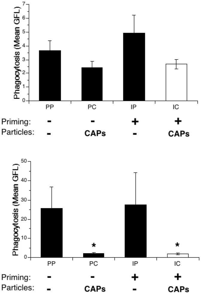
Figure 6.
Ex vivo bacterial uptake in IFN-γ-primed and particle-exposed mice. Mice were exposed to IFN-γ (I) or PBS (P) aerosol for 15 min., then to intranasal PBS (P) or CAPs (C) 3 hours later. BAL was performed 24 hours later and the collected cells were cultured in the presence of heat-inactivated, FITC-labeled S. pneumoniae for 90 minutes at 37°C. After incubation the cells were transferred to ice and labeled with Gr-1 antibody, which binds specifically to PMNs. Flow cytometry followed, measuring green fluorescence (FITC-labeled S. pneumoniae) and gating the cells by their red fluorescence (Gr-1 antibody). Top panel: AMs. Bottom panel: PMNs. Data represent the mean ± SEM of at least 3 independent experiments; * = p<0.05 compared to PP and IP group.

