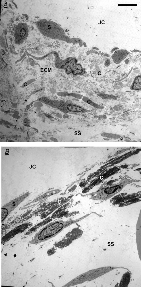
Figure 4. Effect of intra-articular chymopapain on synovial ultrastructure.
A, electron micrograph of control rabbit knee synovium in transverse section B, synovium from a knee treated with 0.1 i.u. intra-articular chymopapain for 20 min. Both panels are ‘Gold’ sections (50–90 nm) of suprapatellar synovium normal to the surface stained with uranyl acetate and lead citrate. JC, joint cavity; S, synovial lining cell; SS, subsynovium; C, collagen bundle; and ECM, extrafibrillar matrix. Note the persistence of collagen fibrils after chymopapain treatment and the increased distinctness of collagen bundles owing to loss of the lightly staining extrafibrillar matrix. Scale bars represent 5 μm.

