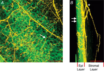
Figure 1. Anatomy of sensory nerve terminals in the rat cornea.
A, flattened Z-stack image of cornea loaded with FM1-43. On the basis of their distinctive morphology, nerve fibres and epithelial cells are artificially coloured in yellow and green, respectively. Arrow indicates nerve terminal residing in the superficial epithelial cell layer. Scale bar, 50 μm. B, same Z-stack as in A but flattened with a 90 deg orientation with respect to A. In this orientation, the thickness of the epithelial cell layer (Epi Layer) is evident. Double arrows indicate surface of the epithelial cell layer. Asterisk indicates subepithelial nerve plexus residing in the stromal layer (collagen layer) of the cornea. Scale bar, 100 μm.

