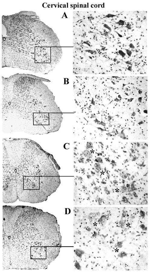
Figure 2. Motor neurons in the cervical spinal cord of G93A mice at early and late stage of disease (cresyl violet staining).
In the cervical spinal cord, many healthy motor neurons with large soma and neuritic processes were identified in the control C57BL/6J mice at (A) 12–13 weeks of age and (B) 19–20 weeks of age. In G93A mice, numerous motor neurons with vacuolization (asterisks) were found at (C) 13 weeks of age and (D) decreased numbers of motor neurons were noted in 17–18 week old mice. Motor neurons of various sizes displayed vacuolization (asterisks). Scale bar on left side is 200 µm, right side is 50 µm.

