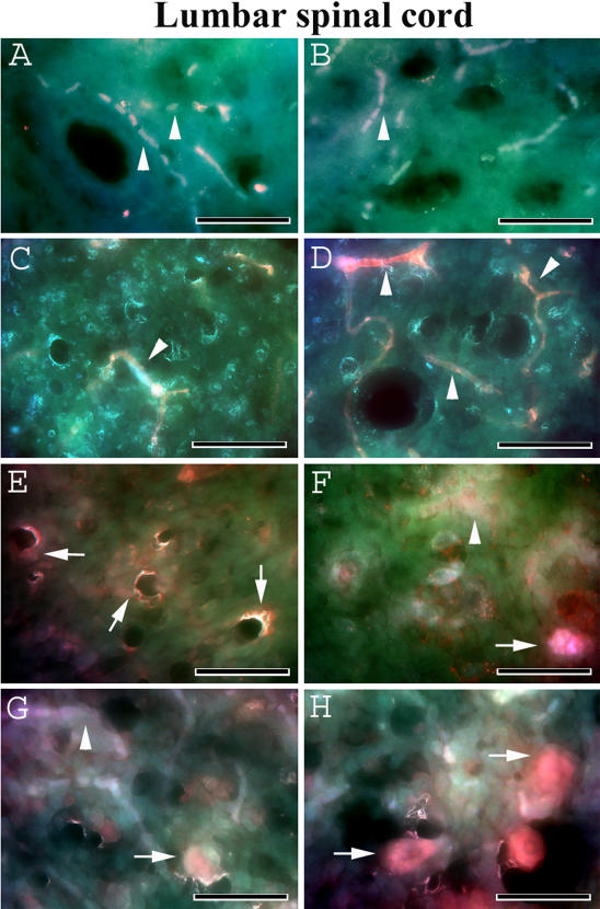
Figure 5. Evans Blue fluorescence in the lumbar spinal cord of G93A mice at early and late stages of disease.
In the lumbar spinal cord, EB dye (red, arrowheads) was determined intravascularly in the control C57BL/6J at (A, B) 12–13 weeks of age and (C, D) 19–20 weeks of age similar to the cervical spinal cord. EB extravasation abnormalities were found in G93A mice at (E, F) 13 weeks of age (red, arrows). (G, H) Significant EB diffusion (red, arrows) into the parenchyma of the lumbar spinal cord from many blood vessels was detected in G93A mice at end-stage of disease (17–18 weeks of age). Arrowheads in F and G indicate vessel permeability. Scale bar in A–H is 25 µm.

