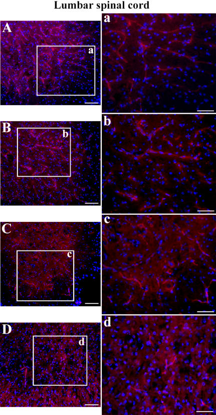
Figure 7. Immunofluorescence staining for laminin in the lumbar spinal cord of G93A mice at early and late stages of disease.
Various laminin-positive vessels (red) were observed in the control C57BL/6J mice at (A) 12–13 weeks of age and (B) 19–20 weeks of age similar to cervical spinal cord results. Fewer blood vessels were labeled in G93A mice at (C) early or (D) end-stage of disease. The nuclei in A–D are shown with DAPI. Scale bar in A, B, C, D is 200 µm; inserts a, b, c, d is 50 µm.

