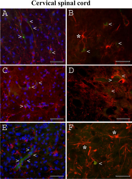
Figure 9. Immunohistochemical staining for endothelial cells (CD146) and astrocytes (GFAP) in the cervical spinal cord of G93A mice at early and late stages of disease.
(A, B) Normal appearance of endothelial cells (green, arrowheads) and delineated astrocytes (red, asterisk) was observed in the control C57BL/6J mice at 19–20 weeks of age. Endothelia (green, arrowheads) surrounding capillaries were partially revealed in G93A mice at (C, D) initial or (E, F) late stages of disease. Note: increased astrocyte activation in the cervical spinal cord (F, asterisks) was detected in G93A mice at late stage of disease. The nuclei in A, C, and E are shown with DAPI. Scale bar in A, C, E is 50 µm; B, D, F is 25 µm.

