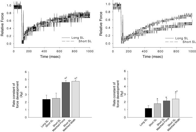
Figure 7. Sarcomere length dependence of rate constant of force development in α-MyHC and β-MyHC cardiac myocytes.
A, normalized force traces fitted with a rising exponential to calculate the rate constant of force development from an α-MyHC myocyte at long SL (upper continuous line) and short SL (lower dashed line). B, normalized force traces fitted with a rising exponential from a β-MyHC myocyte at long SL (lower continuous line) and short SL (upper dashed line). C, rate constants of force development for α-MyHC myocytes at long SL, short SL, short SL with matched force, and short SL with matched width. D, rate constants of force development for β-MyHC myocytes at long SL, short SL, short SL with matched force, and short SL with matched width. *Significantly different from long SL (P < 0.05). †Significantly different from short SL (P < 0.05). Data are means ±s.d.

