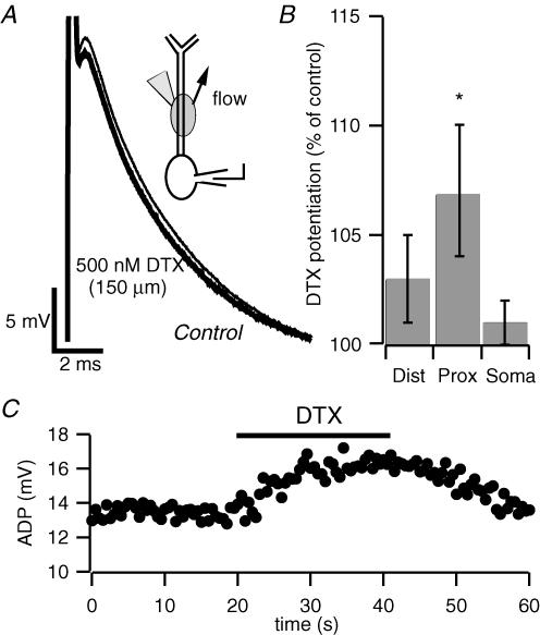
Figure 5. Local application of dendrotoxin to proximal apical dendrites enhances the afterdepolarization.
A, current-clamp recording from the soma of a pyramidal neuron. Focal dendritic application of DTX at 150 μm from soma enhanced the ADP. The inset schematic diagram depicts local application of DTX to apical dendrites. The arrow represents the direction of the superfusion flow. The application pipette contained 500 nm DTX. B, bar chart depicting the potentiation of the somatic ADP by local application of DTX in three different cell compartments. Application of DTX to dendrites 250 μm and beyond (distal) resulted in a slight, non-significant, increase in the size of the ADP (n = 13, P = 0.2); application at 50—200 μm from the soma (proximal) caused an enhancement of the ADP (n = 17, *P < 0.05); application to the soma did not affect the ADP (n = 8). C, time course of the enhancement of the somatic ADP by local application of DTX to the proximal apical dendrite. Action potentials were elicited by 2 ms current injections of 1.1 nA at a frequency of 0.03 Hz from a holding potential of –67 mV. Note the reversibility of the effect upon washout of the toxin.

