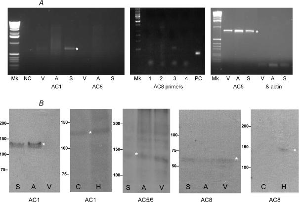
Figure 1. RT-PCR and immuno-blot identify Ca2+ sensitive ACs in SAN cells.
A, RT-PCR of cardiac tissue: SAN (S), atrium (A) and ventricle (V), negative control (NC) and positive control (PC) with consensus primers designed to recognize AC1, AC8, AC5 and β-actin in other mammalian species. A range of different AC8 primers (1–4) were tested in the SAN without effect, despite PC finding the experiment to be effective. B, immuno-blot of cardiac tissue using polyclonal antibodies raised against peptide sequences in AC1, AC5/6 and AC8 common to other mammalian species. Brain extracts, cerebellum (C) and hippocampus (H), were also used to test for AC1 and AC8. Molecular mass markers are shown at the left. The asterisks draw attention to specific DNA or protein bands.

