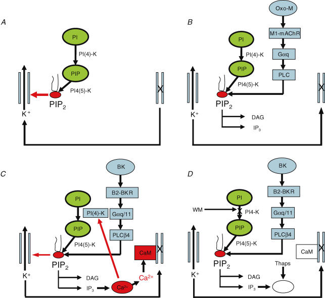
Figure 3. Pathways for M-channel activation and inhibition.
A, channels are normally activated by membrane PIP2. B, muscarinic receptor stimulation closes M-channels by reducing membrane PIP2. C, bradykinin closes channels through IP3-mediated Ca2+ release. D, when Ca2+ release is suppressed and PIP2 synthesis blocked, bradykinin can instead close channels by reducing PIP2. Abbreviations: PI, phosphatidylinositol; PIP, phosphatidylinositol-4-phosphate; PIP2, phosphatidylinositol-4,5-bisphosphate (PI(4,5)P2); PI(4)-K, phosphatidylinositol-4-kinase; PI4(5)-K, phosphatidylinositol-4-phosphate; Oxo-M, oxotremorine-M; M1-mAChR, M1 muscarinic acetylcholine receptor; Gαq, α-subunit of the G proteins Gq; Gαq/11, α-subunit of the G proteins Gq and/or G11; PLC, phospholipase-C; PLCβ4, phospholipase-C β4; DAG, diacylglycerol; IP3, inositol-1,4,5-trisphosphate; BK, bradykinin; B2-BKR, B2-bradykinin receptor; CaM, calmodulin; WM, wortmannin; thaps, thapsigargin. See text for further details. (Adapted from Hughes et al. (2007), with permission.)

