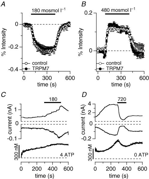
Figure 6. Overexpression of TRPM7 does not alter cell volume regulation in response to osmotic challenges.
A, fluorescent indicator dye imaging of cell volume change of wild-type (^, n = 5) or TRPM7 overexpressing HEK293 cells (•, n = 5) pre-loaded with Fura-2 ester and evalulated at 360 nm excitation. Intact Fura-2-loaded cells were superfused with 180 mosmol l−1 external solution as indicated by the black bar. The concomitant cell swelling was assessed as emission intensity decrease in per cent. Data were normalized to the intensity before application. Error bars indicate s.e.m.B, fluorescent indicator dye imaging of cell volume change of wild-type (^, n = 4) or TRPM7 overexpressing HEK293 cells (•, n = 3) pre-loaded with Fura-2 ester and evaluated at 360 nm. Intact Fura-2-loaded cells were superfused with 480 mosmol l−1 external solution as indicated by the black bar. The concomitant cell shrinkage was assessed as emission intensity increase in per cent. Data were normalized to the intensity before application. Error bars indicate s.e.m.C, combined Fura-2 and patch-clamp experiments. Upper panel shows development of inward and outward currents of an example cell where currents were assessed at −80 mV and +80 mV, respectively. Application of 180 mosmol l−1 as indicated by the bar. Cells were perfused with standard internal caesium glutamate-based solution supplemented with 4 mm Mg·ATP, with 0.9 mm free Mg2+ and 200 μm K5Fura-2. Lower panel shows concomitant intracellular calcium concentration of the same cell. Note that currents are caused by a Cl− conductance, not TRPM7 (n = 4). D, combined Fura-2 and patch-clamp experiments. Upper panel shows development of inward and outward currents of an example cell where currents were assessed at −80 mV and +80 mV, respectively. Application of 720 mosmol l−1 as indicated by the bar (n = 5). Cells were perfused with standard internal caesium glutamate-based solution in the absence of Mg·ATP, with 0.9 mm free Mg2+ and 200 μm K5Fura-2. Lower panel shows concomitant intracellular calcium changes of the same cell.

