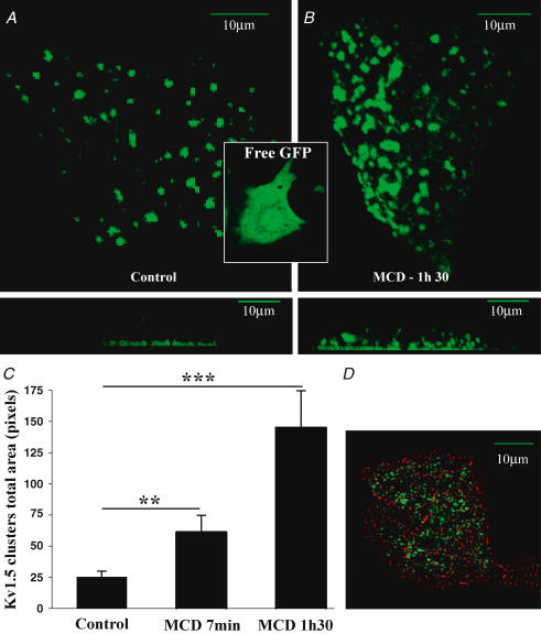
Figure 8. Surface expression of Kv1.5 subunits in neonatal cardiomyocytes.
A, in live cardiomyocytes, transfected GFP-tagged Kv1.5 subunits are clustered at the membrane surface adjacent to the bottom of laminin-coated glass support, as shown in the projection of Z sections in the lower panel. In contrast, GFP alone was homogeneously distributed in cardiomyocytes (inset). B, after the application of 2% MCD, clusters increased in size and were redistributed throughout the plasma membrane. C, bar graphs summarizing changes in cluster size upon MCD exposures; data are from 21 cardiomyocytes in control, and following incubation with 2% MCD for 7 min and 1 h 30 min. **P < 0.01, ***P < 0.001. D, double immunostaining of fixed cardiomyocytes using sarcomeric α-actinin and anti-Kv1.5 antibodies showing that Kv1.5-GFP-transfected cells are cardiomyocytes. A, B and D: scale bars represent 10 μm.

