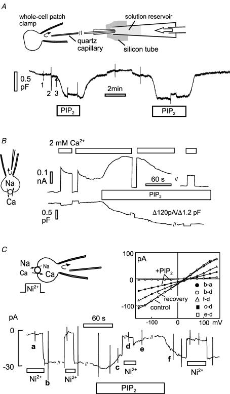
Figure 5. Capacitance and NCX1 current changes associated with perfusion of PIP2 into cells in whole-cell voltage clamp.
A, illustration of pipette perfusion method and the reversible effects of perfusing PIP2 (40 μm) into an A9 cell. Solutions are changed by removing (1) and attaching (2) solution reservoirs (10–50 μl) to a quartz capillary through a silicon sleeve and applying positive air pressure to the reservoir (3). The decrease of capacitance (0.6 pF) corresponds to 5% of the cell capacitance. B, effects of perfusing PIP2 into a BHK cell expressing NCX1. Outward exchange current is activated by applying 2 mm Ca2+ on the extracellular side. With PIP2 perfusion (40 μm), current increases briefly and then decreases over 2 min in parallel with a 12% decrease of membrane capacitance. C, PIP2 inhibition of NCX1 currents with physiological ion gradients. Exchange current is defined by application and removal of 4 mm Ni2+ in the extracellular solution. I–V relations were taken at points marked a–f. Plots are given for subtracted I–Vs at the start of the experiment (b – a and b – d), during perfusion of PIP2 (c – d and e – d), and after removal of PIP2 (f – d).

