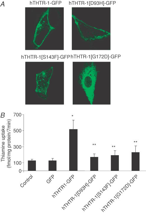
Figure 6. Distribution of individual clinically relevant hTHTR-1–GFP mutations in ARPE-19 cells.
A, representative of confocal lateral (xy) images showing localization of individual mutant constructs in ARPE-19 cells, 24–48 h after transient transfection. B, uptake of [3H]-thiamine in control, GFP, hTHTR-1–GFP, hTHTR-1[D93H]–GFP, hTHTR-1[S143F]–GFP and hTHTR-1[G172D]–GFP transiently expressing ARPE-19 cells. *Significantly different from control (P < 0.01); **no significant difference between mutants and control. Results represents means ±s.e.m. from three separate determinants performed on three different occasions.

