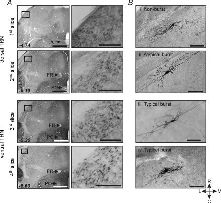
Figure 1. Localization of GAD immunoreactive neurons within TRN.
A, the dorsal to ventral horizontal brain slices (1st slice to 4th slice) are shown to have the GAD immunoreactivity in the TRN indicating the 1st slice that includes the extreme dorsal portion of TRN to the 4th slice include the GABAergic TRN neurons. With lower power objective, the posterior commissure (PC) and fasciculus retroflexus (FR) are used to distinguish the level of each slice. The 1st to 4th slices correspond to −4.74, −5.10, −5.32 and −5.60 mm bregma (Paxinos & Watson, 1986). Scale bars refer to 1 mm and 200 μm. Each rectangle (left) is shown at higher power (right) to illustrate GABA-positive neurons in TRN. B, photomicrographs of different TRN neurons based on their burst discharge characteristics. Four TRN neurons (i–iv) were localized in the 1st to 4th slices. Non-burst neuron (i), atypical burst neuron (ii), and typical burst neuron (iii) have similar morphological characteristics. They have fusiform somas and several primary dendrites that extend from each pole of the cell body and are orientated to the longitudinal axis of the TRN. However, a subpopulation of all three subtype TRN neurons also has somewhat round-shaped soma with multipolar dendrites (iv), suggesting morphological diversity within each subtype. Scale bar, 100 μm. C, caudal; R, rostral; M, medial; L, lateral.

