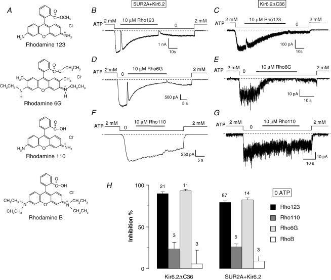
Figure 5. KATP channel inhibition by various rhodamine derivatives.
A, chemical structures of the tested rhodamine derivatives. B–G, currents were recorded in inside-out patches from Xenopus oocytes expressing either SUR2A + Kir6.2 (B, D and F) or Kir6.2ΔC36 (C, E and G). Indicated rhodamine compounds (10 μm) were applied in the absence of nucleotides. H, percentage inhibition by a 10 μm concentration of the indicated compounds of Kir6.2ΔC36 and SUR2A + Kir6.2 channels in the absence of nucleotides. Numbers above bars indicate the number of patches included in the average.

