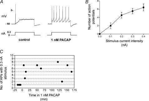
Figure 3. An increased excitability was sustained during bath application of 1 nm PACAP.
A, a recording from the same cell obtained prior to and 5 min after bath application of 1 nm PACAP. Note that the firing pattern elicited with a 1 s depolarizing pulse shifted from phasic to tonic-like during exposure to 1 nm PACAP. B, an excitability curve demonstrating that during continued exposure to 1 nm PACAP, the number of action potentials elicited by 1 s depolarizing current pulses increased with increased stimulus strength (averaged data from 9 cells that were evaluated at different times over a 5–166 min exposure to PACAP). C, the number of action potentials produced by a 0.3 nA stimulus in different cells are plotted as a function of time in 1 nm PACAP. The results show that neuronal excitability remained elevated during the sustained exposure to PACAP.

