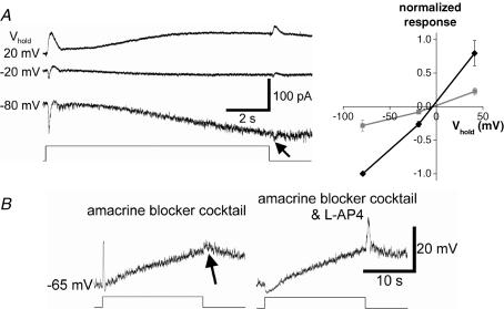
Figure 8. Light-induced synaptic inputs are also driven by ON and OFF bipolar cells.
A, in the presence of pharmacological blockade of amacrine cell inputs (see Methods), the extrinsic light response reversed near 0 mV, implicating a cationic current. Left, current recordings at several holding potentials; the traces are averages of several responses. Note the small response at light OFF (arrow) in addition to the larger ON response. Light intensity =−2 log I. Right, baseline-subtracted I–V plot of peak currents evoked at light ON (black) and light OFF (grey), showing that these currents reversed near 0 mV and thus were mediated by an increase in cationic conductance. This plot was averaged from all five cells tested, with each cell's ON response amplitude at −80 mV normalized to −1. Error bars indicate s.e.m. values. B, voltage responses of another ipRGC in the same amacrine blocker cocktail similarly revealed components at light OFF (arrow) as well as at ON. The ON depolarization was selectively abolished by further addition of l-AP4 while the OFF response was enhanced, indicating that the OFF depolarization was generated by OFF bipolar cells rather than by the surround response of ON bipolar cells. Light intensity =−2.5 log I.

