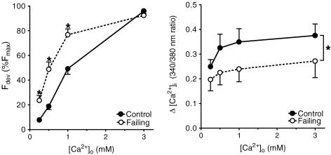
Figure 4. Effect of external [Ca2+]o on developed force (left panel, normalized to Fmax) and the amplitude of intracellular [Ca2+]i (right panel) at 0.5 Hz and 27°C from control (n= 10) and failing (n= 10) right ventricular trabeculae.
A leftward shift in the force–[Ca2+]o relation in the failing group indicates an increased sensitivity of the cardiomyocytes to external calcium, which is accompanied by a decreased amplitude of [Ca2+]i as compared with control preparations. Force and amplitude of [Ca2+]i increased with [Ca2+]o in both groups. Values are expressed as means ±s.e.m.*P < 0.05 versus control group.

