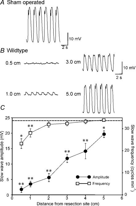
Figure 1. Disruption of pacemaker activity in the small intestine 5 h after intestinal resection.
A and B, typical intracellular electrical activity (slow waves) recorded from the circular muscle of the small intestine of a sham-operated mouse (A) and at various distances (0.5–5 cm) from the site of an intestinal anastomosis in a BALB/c mouse (B). Slow-wave amplitude was dramatically reduced near the site of anastomosis and increased in amplitude as a function of distance from the resection site. C, summary of the changes in slow-wave amplitude (•) and frequency (□) as a function of distance from the site of anastomosis (n= 13 animals). The data are compared to the amplitude of slow waves in sham-operated animals (dashed line; n= 8 animals). Slow-wave amplitude was significantly reduced at all sites tested after surgery compared with sham-operated animals. Frequency was reduced only at 0.5 and 1 cm from the site of the anastomosis (*P < 0.05, **P < 0.01). Values in the amplitude and frequency of slow waves were significantly different from each other within the group of the control operation model (both P < 0.001, by ANOVA).

